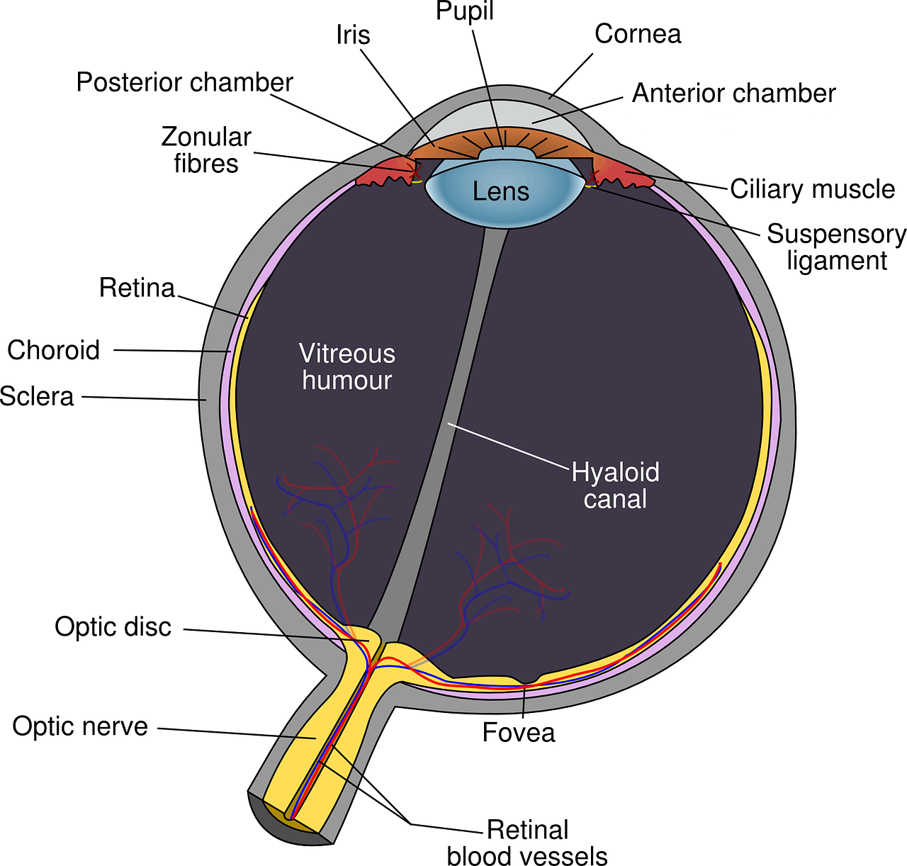
Eye Anatomy
Anterior Chamber, Fluid-filled space inside the eye that is located between the iris and the innermost corneal surface (endothelium).
Choroid, Vascular (major blood vessel) layer of the eye lying between the retina and the sclera. The choroid provides nourishment to outer layers of the retina.
Ciliary Muscle, is a ring of smooth muscle in the eye’s vascular layer that controls viewing objects at varying distances and regulates the flow of aqueous humour into Schlemm’s canal.
Cornea, front part of the eye that is transparent and covers the iris, pupil, and anterior chamber and provides most of an eye’s optical power.
Fovea, pit in the central part of the macula that produces sharp vision.

Hyaloid Canal,is a small transparent canal running through the vitreous body from the optic nerve disc to the lens that facilitate changes in the volume of the lens.
Iris, is the circular structure in the eye, responsible for controlling the diameter and size of the pupil and the amount of light reaching the retina. The Iris also helps give color to the eye.
Lens, transparent, convex structure in the eye that helps to refract light to be focused on the retina by changing shape.
Optic Disc, is the point of exit for ganglion cell axons leaving the eye. It is located at the head the optic nerve.
Optic Nerve, is one of the main nerves that transmits visual information from the retina to the brain
Posterior Chamber, is a narrow space behind the iris and in front of the lens.
Pupil, the round, centered portion of the eye, which opens and closes to regulate the amount of light the retina receives.
Retina, light-sensitive layer of tissue within the inner coat of the eye. The retina converts images from the eye’s optical system into electrical impulses that are sent along the optic nerve to the brain.
Sclera, the protective outer coating of the eyeball that forms the visible white of the eye.
Suspensory ligament, a ring of fibrous tissue that attaches to the lens and holds it in place.
